 PEER-TO-PEER
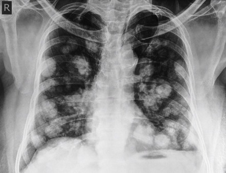
This illustrated case report displays a 54-year-old man diagnosed with metastatic embryonal carcinoma presenting with cannonball pulmonary lesions. The patient’s initial symptoms, diagnostic workup, and treatment plan highlight the importance of recognizing this radiographic pattern and its implications for potential underlying malignancies.
Key Points:
- A 54-year-old man with mild intermittent asthma presented with worsening shortness of breath and cough over 2 months.
- Chest imaging revealed numerous round opacities in both lungs, known as “cannonball” pulmonary lesions.
- Laboratory tests showed elevated lactate dehydrogenase and beta human chorionic gonadotropin levels.
- Ultrasonography discovered a right testicular mass, and biopsy confirmed metastatic embryonal carcinoma.
- The patient received platinum-based chemotherapy followed by salvage chemotherapy.
- Cannonball pulmonary lesions typically indicate hematogenous cancer spread but can rarely occur in infectious or autoimmune conditions.
- The patient is being evaluated for autologous stem-cell transplantation.
Pulmonary metastases are usually asymptomatic or can present with hemoptysis, dyspnea, and pneumothorax. Symptoms more commonly arise from the primary tumor, extrapulmonary metastates or systemic effects. Pulmonary thrombotic microangiopathy is an exception, causing hypoxemia, pulmonary hypertension, cor pulmonale, rapid decline and death. Gastric carcinoma is the most common primary. (Radiopaedia)
More on Diagnostic Radiology