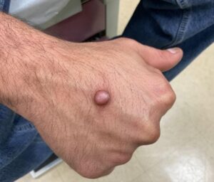
Implications for Diagnosis and Treatment of Cutaneous Focal Mucinosis in Dermatological Practice

Cutaneous focal mucinosis represents a unique clinical entity characterized by the localized accumulation of mucin within the dermis. First identified by Johnson and Helwig in 1966, this condition has been further delineated through subsequent research to distinguish solitary lesions from those associated with systemic diseases. The condition’s benign nature, combined with its distinct histopathological features, underscores the importance of accurate diagnosis and appropriate management strategies for dermatologists. This summary distills the key findings and clinical recommendations to aid physicians in the identification, diagnosis, and treatment of cutaneous focal mucinosis.
Key Points:
- Epidemiology and Presentation: Cutaneous focal mucinosis typically presents as solitary, flesh-colored to white or red, asymptomatic, dome-shaped papules or nodules, with a slight male predilection and an age range of presentation from 29 to 60 years.
- Differential Diagnosis: The clinical differential diagnosis includes dermatofibroma, neurofibroma, basal cell carcinoma, and several other skin conditions, necessitating thorough examination and history-taking.
- Histopathological Confirmation: Diagnosis requires biopsy, with hematoxylin and eosin staining revealing unencapsulated mucin in the dermis. Mucin-specific stains further confirm the diagnosis.
- Treatment Approach: Simple local excision is the treatment of choice, noted for its effectiveness and low recurrence rate.
- Association with Systemic Diseases: Multiple lesions may indicate underlying systemic conditions, including scleroderma and thyroid disease, warranting a comprehensive evaluation.
- Importance of Comprehensive Evaluation: A detailed family history and laboratory evaluation, including tests for TSH, thyroid antibodies, and antinuclear antibody, among others, are recommended for patients presenting with multiple lesions.

The prevalence of mucinosis in dermatology has led to advancements in molecular diagnostics, with studies showing that specific genetic markers may predict susceptibility to related conditions. For instance, research has identified genetic mutations associated with mucin accumulation in conditions like scleromyxedema.
More in Dermatology
