 DIET & NUTRITION
  PATIENT EDUCATION
  OBESITY/WEIGHT MANAGEMENT
  EXERCISE/TRAINING
  LEGAL MATTERS
  GUIDELINES/RECOMMENDATIONS
 Distinguishing Dermatological Diagnoses: A Case of Progressive Skin Discoloration
A 77-year-old male patient presents for evaluation, revealing a hyperpigmented macule on his right cheek. This lesion has been gradually enlarging and darkening, as observed by his wife over the past year. Dermoscopic examination offers key insights but leaves room for differential diagnosis.
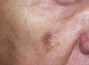
Considerations include:
- Macular seborrheic keratosis: A benign skin growth, typically waxy and raised, often found on sun-exposed skin.
- Pigmented basal cell carcinoma: A slow-growing skin cancer, often arising in sun-exposed areas, and appearing as a pink, brown, black, or translucent bump.
- Lentigo maligna: A type of in situ melanoma found primarily on chronically sun-exposed skin. It usually begins as a macular skin discoloration that can range from light to dark brown, sometimes with pink, red, or white hues.
- Superficial spreading melanoma: This common type of melanoma often presents as a flat or slightly raised discolored patch with irregular borders, typically resulting from intense UV exposure.
Based on these findings, what is your diagnosis?
Dermatology Latest Posts
- Quality of Life in Young Adults With Acne
- Digital Patient Navigation Platform Fosters Engagement Between Physicans and Patients with Melanoma
- Radiofrequency Microneedling: Postmarketing Data Reveals Adverse Effects
- FDA Approves OTC Switch for Galderma’s Differin Epiduo Acne Gel
Did You Know?
Lentigo maligna, an in situ subtype of melanoma, affects primarily males between the ages of 60 and 80 years, with over 50% of cases developing on the cheeks.